Jessica Alber developing non-invasive prevention scanning
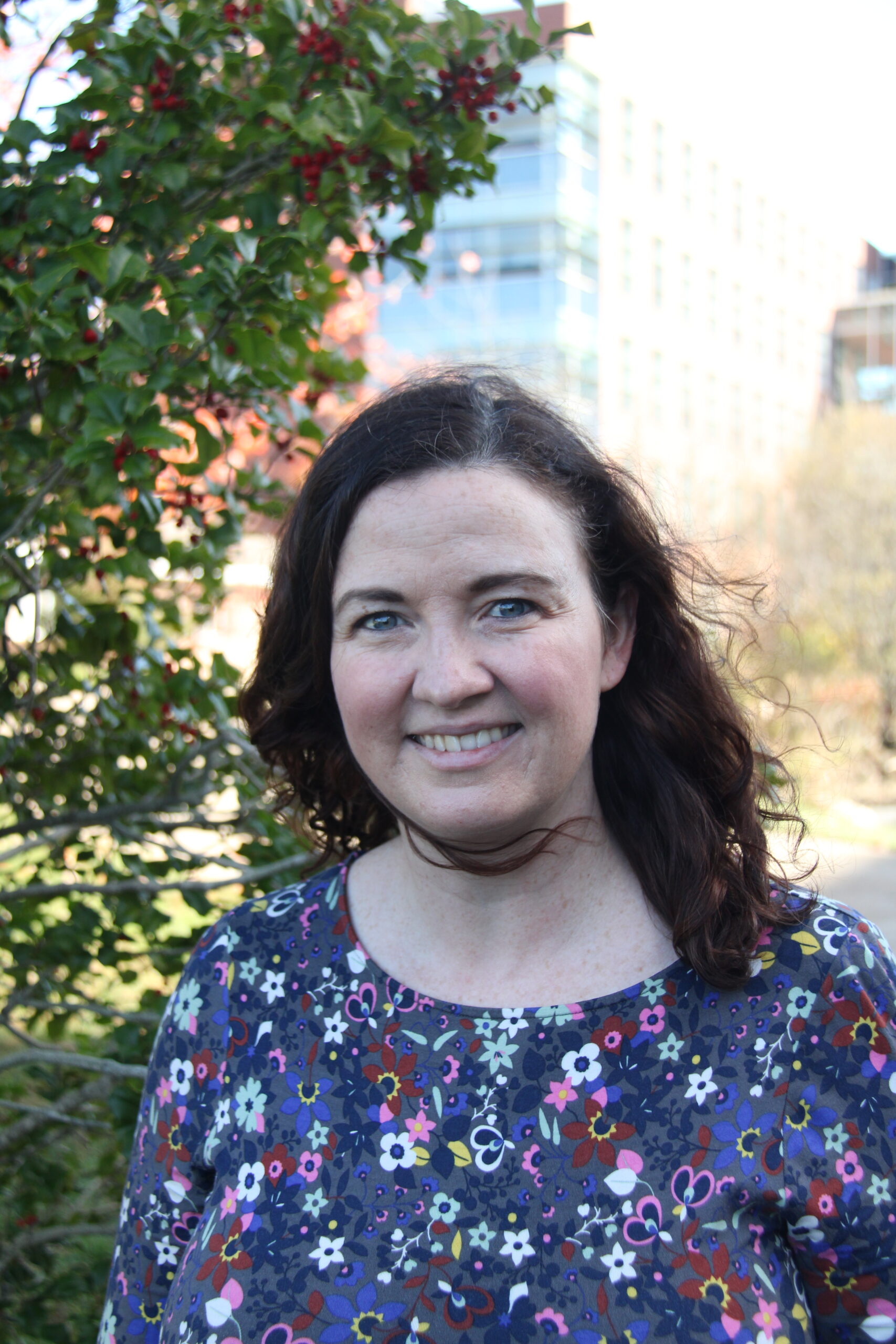
Assistant research professor Jessica Alber received a grant for her work with Alzheimer’s disease. PHOTO CREDIT: Nick Pierson | Contributing Photographer
Jessica Alber, an assistant research professor at the University of Rhode Island’s George and Anne Ryan Institute for Neuroscience, just received a five-year, $10.3 million grant to further develop a screening technique that detects Alzheimer’s disease before symptoms appear.
After receiving funding from the National Institute of Health (NIH), the assistant professor of biomedical and pharmaceutical sciences is looking to change the way doctors diagnose and possibly treat Alzheimer’s disease.
Alber’s project, “Longitudinal validation of retinal biomarkers against cerebral imaging in preclinical Alzheimer’s disease,” could implement a means of early detection that is “low-cost, non-invasive… and more accessible to people.”
While affordability and accessibility make her research alluring, Alber believes that the main benefit of this screening tool is the connection between early intervention and treatment success.
Currently, there are no treatments that impede the progression of Alzheimer’s disease, according to Alber. However, with growing research into how combinations of lifestyle modification and drug treatment impact Alzheimer’s disease, Alber believes that early detection could be a key to eventual success in treatment.
Alzheimer’s disease is a type of dementia characterized by memory impairment, functional loss and a decline in activities of daily living. The disease also requires an official diagnosis.
“By the time someone shows up with memory lapses or loss of daily functioning… it’s too late,” Alber said. “We really need to catch [Alzheimer’s disease] before any symptoms present themselves.”
The NIH grant facilitates Alber’s goal by funding the second phase of the Atlas Retinal Imaging in Alzheimer’s Study (ARIAS), which was led by Peter Snyder, URI’s vice president for research and economic development.
Launched in 2020, the ARIAS study focused on using “structural, anatomic and functional imaging of the retina” to identify changes directly related to Alzheimer’s disease, according to Snyder.
By creating a reference database using these identified pathological changes, Snyder explained that he developed biomarkers of Alzheimer’s prospect and progression, allowing doctors to spot the disease before cognitive symptoms arise.
He said that the biomarkers found in his study could be used by optometrists in the future, and people who are at genetic risk for Alzheimer’s disease could check for evidence of the disease each time they go for an eye checkup.
The clinical trials in Snyder’s ARIAS study used a non-invasive imaging technology called optical coherence tomography (OCT) to capture high-resolution images of the retina. Snyder explained that by directing lasers at the retina and recording the light reflected back into the camera, OCT can spot the buildup of a protein called amyloid plaque. This plaque, although invisible to the naked eye, is “a distinct marker of [Alzheimer’s] disease itself.”
Now that Snyder discovered which changes in the retina would be good candidates to use in an eye doctor’s office, Alber is focusing on validating their findings.
The main focus of Alber’s study, named ARIAS 2, is to take these observations from Phase I and compare them to what she calls a “ground truth.” This involves looking at physical changes in the brain to determine if they reflect a neurodegenerative process or changing proteins indicative of Alzheimer’s disease – as hypothesized in the original ARIAS study.
“Validation is the next logical step,” Alber said. “We can’t say something works in syntax unless we’ve tested it.”
She has clinical sites at the Butler Hospital Memory and Aging Program in Providence, Washington University at St. Louis School of Medicine and the University of North Texas Health Sciences Center at Fort Worth. Butler Hospital was a study site for the first ARIAS study and is an affiliate of Brown University, where she once worked as an assistant professor.
The first step of ARIAS 2 involves looking at people’s brains and eyes at the same time, according to Alber. This is done through magnetic resonance imaging (MRI), where researchers study the actual volume and structure of the brain.
Researchers perform these techniques and scans on individuals with a predisposition to Alzheimer’s disease and then see how the results differ from “normal” brains and eyes. They have people fill out questionnaires about their day-to-day and cognitive functioning and then perform retina imaging and MRIs. Using these techniques, Aber and her collaborators can demonstrate the validity of their findings in a non-invasive manner.
As well as verifying the previously identified retinal imaging markers, ARIAS 2 will build upon Snyder’s work by concentrating on another possible detector of Alzheimer’s disease: blood plasma.
Blood tests can be done by primary care physicians, are low-cost and don’t require the individual to go through brain imaging or exposure to radiation, according to Snyder.
He said that clinics currently detect Alzheimer’s disease through invasive and costly methods. Because of this, not only is the disease caught too late for a chance at successful treatment, but individuals are subject to radiation and side effects of medication.
Clinicians use treatments such as positron emission tomography (PET) and lumbar puncture to spot the protein buildup of amyloid and tau plaque. While these methods are effective at detecting those two key proteins, Alber seeks to find a way around the “18-gauge needle [used] in PET scans.”
Clinicians recommend that those who are at high risk of developing Alzheimer’s disease, such as individuals who smoke or are diabetic, are recommended to exercise and consider a Mediterranean diet, according to Snyder. Although medications for the disease “don’t work very well and have risks,” he believes that future drugs could work better if they are introduced before symptoms appear.
Alber’s plans past ARIAS 2 concentrate on integrating their retinal screening techniques into optometrist visits. Alber and Snyder both asserted that since many people visit the eye doctors yearly anyway, this integration would not be a significant change in routine.
Since funding for Alzheimer’s research has opened up over the last five years, Alber explained the importance as a researcher of having the funds and resources needed to “make a contribution to society at large in some way.” Alber stated that, although she is fairly early in her career, she has been building towards an opportunity like this.
Alber believes that this NIH grant will help her make a positive impact on people’s lives. Three out of four of her grandparents died of Alzheimer’s disease, and Alber hopes to implement the ARIAS retinal screening technique “if not in [her] lifetime, but [her] daughter’s.”
By creating an inexpensive and noninvasive means of detecting Alzheimer’s disease up to two decades before symptoms appear, Alber’s research has the possibility to change how clinicians treat the disease.
“We don’t need help figuring out who has Azheimer’s disease when they’re symptomatic, Snyder said. “We need to find people 20 years before that. And that’s what we’re going to do.”